Brain Imaging: A Guide for Clinicians
Synopsis
Brain Imaging: A Guide for Clinicians is designed to provide a foundation of information necessary to those wishing to integrate brain imaging into their practice, or to those that currently review brain scans but have minimal formal training in neuroimaging. The guide covers a range of topics important to those using brain imaging, such as the strengths and weaknesses of the many different techniques currently available, the factors that may influence the
use of imaging data, common pitfalls or artifacts that may be misleading to the clinician, the most appropriate techniques to use given a specific clinical question or condition, how to interpret information presented on a brain image, and also how many pathological conditions appear on a variety of brain
scanning techniques or sequences. This guide also provides detailed information regarding the identification of primary brain regions, anatomical structures, systems or pathways using both two-dimensional and three-dimensional imaging techniques. A brain atlas is included using both CT and MRI sequences to facilitate the reader's ability to identify most primary brain structures. A novel color-coded system is used throughout this guide to assist the reader in identifying slice locations and
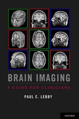
orientations. Images with green borders are displayed in the axial plane, with the slice location being shown on other orthogonal image planes by a green line. Similarly, images with a red border are displayed in the coronal plane and those with a blue border are displayed using a sagittal plane;
red and blue reference lines are displayed on orthogonal slices to identify the slice location. The crosshairs formed by the color-coded reference lines optimize the reader's ability to identify primary anatomical structures or pathological markers and processes.
This book is written in a manner to progress from a general description of the clinical use of brain images and the interpretation of brain scans, to more complex chapters involving neuroanatomy and imaging technology. Real life examples of clinical cases are integrated into all chapters of this guide. Brain Imaging: A Guide for Clinicians provides hundreds of images derived from traumatic and non-traumatic pathologies to provide the reader with examples of conditions most often seen
in the clinic. PEARL-PERIL sections outline critical information for the clinician, along with many tables and charts designed to provide general information required when interpreting brain images.
Publisher information
- Publisher: Oxford University Press Inc
- ISBN: 9780199764679
- Number of pages: 432
- Dimensions: 283 x 222 x 27 mm
- Weight: 1708g
Customer Reviews
You may also like...